

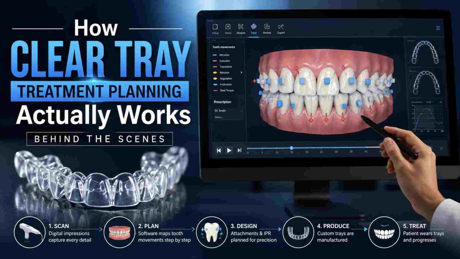
Patients see two things: a scan appointment and, weeks later, a box of trays in numbered order. Everything that happens in between, the part that actually determines whether treatment finishes on schedule and produces the result that was promised, happens somewhere a patient never sees. Understanding that hidden middle stretch matters as much for practices as it does for the people sitting in the chair.
It Starts With a Complete Digital Impression
The process begins the same way nearly every modern workflow does: with a 3D scan that captures the full geometry of a patient’s teeth, gum line, and bite relationship. Unlike a traditional impression, a digital scan can be reviewed immediately for completeness, which means a poor capture gets caught and redone on the spot rather than discovered days later when a lab calls back asking for a remake. That immediacy alone removes a category of delay that used to be routine in earlier workflows.
What the Scan Actually Captures
A good scan isn’t just a snapshot of tooth position. It captures enough detail to model root position estimates, contact points between teeth, and the full occlusal relationship, all of which feed directly into how the software predicts movement later in the process. Skimping on scan quality at this stage tends to surface as planning problems much further down the line, often in ways that aren’t obvious until the simulation stage flags an inconsistency the original scan should have caught.
Mapping the Full Course of Movement
Once the scan is captured, the real planning work begins, and this is where most of the actual decision-making happens.
Setting the Final Position First
Rather than planning movement step by step, most modern workflows start from the end: the clinician defines the desired final tooth position first, then the software works backward to map a series of incremental movements that get from the starting position to that endpoint. Each increment becomes one tray in the sequence, with the total number of trays depending on how much movement is needed and how finely that movement gets broken into individual steps.
Where Attachments Come Into Play
Certain types of movement, particularly rotation or extrusion, need more than tray pressure alone to execute reliably. Small tooth-colored attachments get planned into specific positions on certain teeth, giving the tray something more precise to grip during those harder movements. Attachment placement isn’t guesswork either; it’s planned digitally alongside the rest of the case, based on which movements each tooth needs to make. Getting attachment position right matters as much as getting tooth position right, since a poorly placed attachment can undermine an otherwise well-planned movement sequence.
Running the Simulation Before Anything Gets Built
Before a single tray gets manufactured, the full treatment plan runs as a simulation that shows projected tooth movement from start to finish.
Why This Step Matters So Much
This simulation is the clinician’s real opportunity to catch problems before they become physical trays sitting in a box. A movement that looks unrealistic, a sequence that seems too aggressive, or a final position that doesn’t account for a bite consideration can all be flagged and revised here, while a correction still only costs a few minutes of review. Once trays are manufactured and shipped, the same correction costs a refinement scan and a delay in treatment, which is exactly the kind of setback careful review at this stage is designed to prevent.
Translating the Plan Into Physical Trays
Once the simulation is approved, the digital plan gets translated into a manufacturing file for each individual tray in the sequence.
From Digital Model to Physical Tray
Each stage of projected movement becomes its own 3D printed model, over which a clear thermoplastic material is formed to create the actual tray a patient will wear. The precision of that translation step matters enormously, since a tray that doesn’t fit the digital model it was built from won’t deliver the exact force the simulation predicted, undermining the whole plan before treatment even starts.
Why This Process Reduces Mid-Treatment Surprises
The entire point of building and reviewing a digital simulation before manufacturing anything is to catch what would otherwise be discovered mid-treatment, after a patient has already started wearing trays.
Fewer Refinement Cycles Down the Line
A well-planned, thoroughly simulated case tends to need fewer refinement scans partway through treatment, since most of the potential issues were already caught and corrected during the simulation review. Digital aligner orthodontics gives practices the ability to front-load that scrutiny, catching problems on a screen instead of discovering them on a patient several months into treatment.
A Better Conversation With Patients
This behind-the-scenes process also gives practices something valuable to share with patients directly: a visual simulation of their own projected results. Showing that simulation during the initial consultation tends to set realistic expectations and build confidence in the plan before a patient ever commits to treatment.
Conclusion
Everything that makes clear tray treatment predictable happens long before a patient receives their first tray, in the scanning, mapping, and simulation stages most patients never see. Getting that hidden process right is what separates a treatment plan that finishes on schedule from one that needs constant correction along the way.